Free T4 and Free T3 Normal Ranges in pmol/L: A US Reader's Guide
Many US labs report free T4 and free T3 in pg/mL or ng/dL, but international results arrive in pmol/L. Here is how to read both, what the ranges mean, and when trends matter more than units.
Medical disclaimer: The information in this article is for educational and informational purposes only. It does not constitute medical advice, diagnosis, or treatment. Lab results and reference ranges vary by individual, lab, age, sex, and health history. Always consult a qualified healthcare provider before making any decisions about your health, medications, supplements, or lab testing. LabHealthCharts is a data visualization tool — it organizes and displays your lab data, it does not interpret your results or provide medical guidance.
Roughly 20 million Americans have some form of thyroid disease, and many more receive thyroid panels as part of routine physicals. But when a result arrives in pmol/L instead of the pg/mL or ng/dL printed on a standard US report, even people who follow their labs closely can feel stuck. The number looks wrong. The reference range seems different. The trend feels unreadable.
This guide explains what free T4 and free T3 actually measure, what both unit systems mean, how to convert between them, and why the direction of those numbers across multiple draws often tells you more than any single result in any unit.
What free T4 and free T3 actually are
The thyroid gland sits at the base of the throat and produces two main hormones: thyroxine (T4) and triiodothyronine (T3). T4 is produced in larger quantities and acts mainly as a storage and transport form. Most of it travels through the bloodstream bound to carrier proteins (primarily thyroxine-binding globulin, or TBG), which makes it inactive. The small fraction that is unbound is called free T4, abbreviated fT4 or FT4. That free portion is what your lab actually measures.
Free T3 (fT3 or FT3) is the biologically active form. Most T3 in circulation is converted from T4 in peripheral tissues, particularly the liver and kidneys, rather than secreted directly by the thyroid. Once T3 enters a cell, it binds to receptors in the nucleus and switches gene expression on or off, affecting metabolic rate, heart rate, body temperature, and dozens of other processes. In plain terms: T4 is what your thyroid ships out, and T3 is what your tissues actually use.
Because both markers float in the same panel, a pattern across them often reveals things neither number alone can show. A post on this site covers the free T3/free T4 ratio in depth; this article focuses specifically on the units, ranges, and how to read results reported in pmol/L.
Why results show up in pmol/L instead of pg/mL
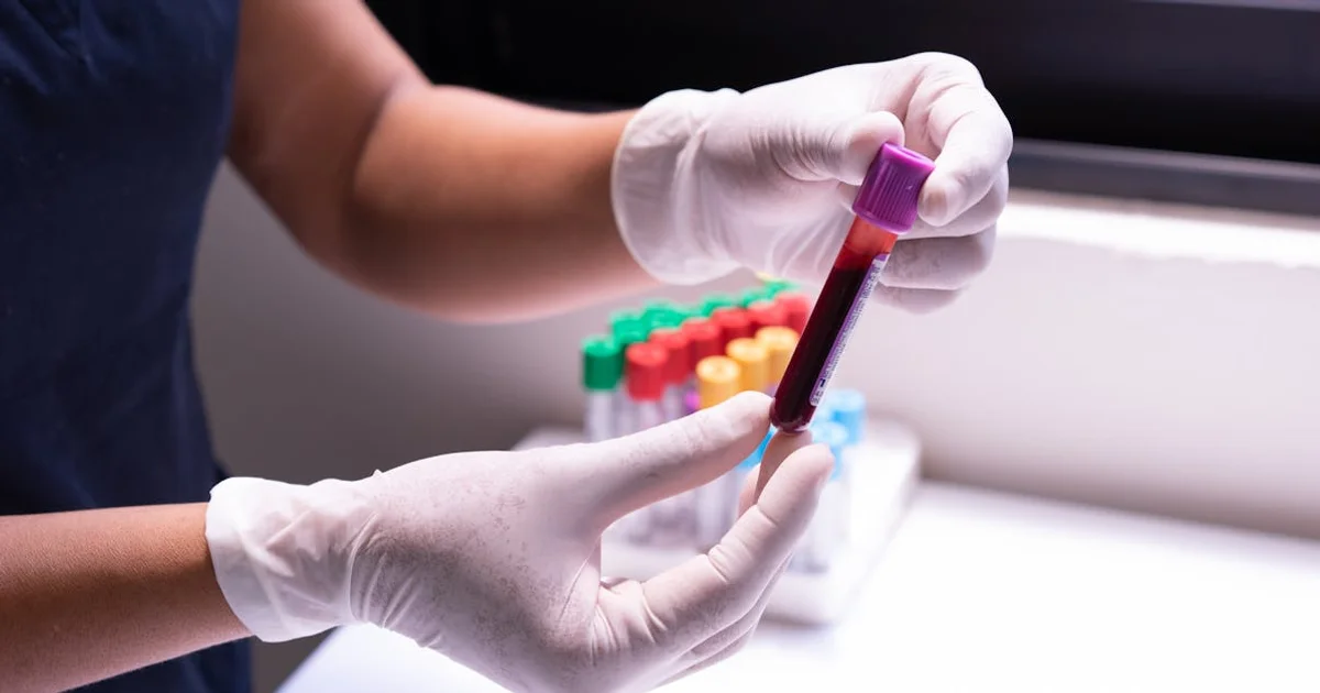
Most US commercial labs (Quest Diagnostics, LabCorp) report free T4 in ng/dL (nanograms per deciliter) and free T3 in pg/mL (picograms per milliliter). International labs, Canadian labs, and some academic medical centers use SI units: pmol/L (picomoles per liter). When you receive results from a lab outside the US, order labs through a service using SI conventions, or look up reference values from a non-US source online, you will see pmol/L.
Neither system is more accurate. They measure the same quantity; only the scale differs. A mole is a count of molecules (6.02 × 10²³ of them), and expressing concentration in moles per liter gives a unit that does not depend on the molecular weight of the substance being measured. Nanograms and picograms are weight-based, so they do depend on molecular weight. Both are precise as long as you stay within one system and use the correct reference range for that system.
Free T4 normal range: ng/dL vs pmol/L
For free T4, the commonly cited reference range at major US clinical laboratories is approximately 0.8–1.8 ng/dL. Converted to SI units, this corresponds to roughly 10–23 pmol/L. The conversion factor is straightforward: multiply ng/dL by 12.87 to get pmol/L (or divide pmol/L by 12.87 to get ng/dL). This factor reflects the molecular weight of thyroxine (776.9 g/mol).
Free T4 reference ranges by unit system (typical values — confirm with your lab's specific reference interval)
| Unit | Typical lower limit | Typical upper limit | Conversion from ng/dL |
|---|---|---|---|
| ng/dL (US labs) | 0.8 | 1.8 | — |
| pmol/L (international / SI) | 10.3 | 23.2 | multiply ng/dL × 12.87 |
| ng/L (less common) | 8 | 18 | equivalent to pg/mL × 10 |
These figures align with ranges published in major endocrinology literature. A large reference study in Clinical Chemistry and guidance from the American Thyroid Association both note that reference intervals differ by immunoassay platform and laboratory method, which is why the range printed on your specific report is the one to use for interpreting your result. A value of 15 pmol/L is solidly mid-range; a value of 9.5 pmol/L sits below the typical lower limit; a value of 25 pmol/L exceeds the typical upper limit. Absolute placement matters, but so does where it was six months ago.
Free T3 normal range: pg/mL vs pmol/L
Free T3 is reported in pg/mL (picograms per milliliter) by most US labs, with a typical reference range of approximately 2.3–4.2 pg/mL. In SI units, this translates to roughly 3.5–6.5 pmol/L. The conversion factor is approximately 1.54 (multiply pg/mL by 1.54 to get pmol/L), reflecting the molecular weight of triiodothyronine (650.9 g/mol).
Free T3 reference ranges by unit system (typical values — confirm with your lab's specific reference interval)
| Unit | Typical lower limit | Typical upper limit | Conversion from pg/mL |
|---|---|---|---|
| pg/mL (US labs) | 2.3 | 4.2 | — |
| pmol/L (international / SI) | 3.5 | 6.5 | multiply pg/mL × 1.54 |
| ng/dL (older notation) | 0.23 | 0.42 | divide pg/mL by 10 |
The GSC queries arriving at this site include searches for "free T3 normal range pmol/L" and "free triiodothyronine FT3 normal range," which suggests many readers encounter results in these units without a clear conversion anchor. A value of 4.8 pmol/L, for example, converts to approximately 3.1 pg/mL, which sits within the typical range. A value of 7.2 pmol/L (approximately 4.7 pg/mL) would be above the upper limit for most lab platforms. Context from the specific lab report matters more than any online table.
The broader scientific evidence on what free T3 measures, and how it behaves differently from free T4, is covered in the site's free T3 explainer and free T4 explainer. Those posts are worth reading alongside this one if you want the full clinical picture.
What actually shifts these values — and why one draw is not enough
Several factors move free T4 and free T3 independently of thyroid disease. Binding protein concentrations change with oral contraceptive use, pregnancy, liver disease, and certain medications, which is one reason the free (unbound) fractions are measured rather than total T4 or total T3 in most modern panels. Even within the free fractions, assay-to-assay variability exists: a 2019 analysis in Thyroid found that free T4 values for the same sample differed by as much as 30% across immunoassay platforms. That variance alone can push a value across a reference range boundary without any change in the patient's actual thyroid status.
Illness, caloric restriction, and acute stress can all suppress free T3 temporarily, a pattern sometimes called non-thyroidal illness syndrome or euthyroid sick syndrome. In these cases, TSH often remains normal, free T4 may be unchanged or mildly low, and free T3 drops because peripheral conversion of T4 to T3 is suppressed as a metabolic adaptation. Recovering from surgery, an acute infection, or significant caloric deficit can temporarily produce a free T3 result in pmol/L that looks alarming out of context, but resolves as the underlying situation improves.
On thyroid hormone replacement therapy (levothyroxine), free T4 tends to normalize first, while free T3 conversion depends on peripheral tissue and varies between individuals. A study in The Journal of Clinical Endocrinology & Metabolism documented that some patients on levothyroxine maintain TSH and free T4 within range while free T3 remains lower than in people with intact thyroid function, a finding that has contributed to ongoing clinical debate about combination T3/T4 therapy. Whether your free T3 on therapy is trending toward or away from the lower limit of range over successive draws is a more useful question than where a single number falls.
The "optimal" range debate: what the evidence actually says
Within the reference range, functional medicine practitioners and some endocrinologists debate whether "optimal" differs from "normal." For free T4, there is no strong clinical consensus that a mid-range value (say, 15 pmol/L versus 18 pmol/L) produces measurably different symptoms in people without thyroid disease. For free T3, the picture is more contested.
Some clinicians observe that patients report better symptom resolution at free T3 levels in the upper half of the reference range. A systematic review in Frontiers in Endocrinology noted significant variation in patient-reported wellbeing at TSH levels that guidelines consider adequate, suggesting that TSH alone does not fully capture treatment response. Free T3 may provide additional information in that gap. The mainstream clinical position, supported by major endocrine societies, is that TSH within range is sufficient for most patients; the functional medicine position holds that free T3 in the upper third of range correlates with better outcomes for some. Both positions cite real data, and neither is settled.
For readers monitoring their own results: note where your free T3 and free T4 sit within their respective ranges, not just whether they are flagged high or low. A free T3 of 3.6 pmol/L (low-normal) in a person who was previously at 5.2 pmol/L is a different clinical situation than a first-ever result of 3.6 pmol/L with no history. That longitudinal context is something a single result printed on a PDF cannot show you.
How free T4 and free T3 fit the broader thyroid picture
These two markers do not exist in isolation. TSH (thyroid-stimulating hormone) is the pituitary signal that drives thyroid output and is typically the first marker ordered. When TSH is abnormal, free T4 is added to clarify whether the thyroid is producing adequate hormone. Free T3 may be added when there is a discrepancy between TSH and free T4, or when a patient on levothyroxine reports persistent symptoms despite a normalized TSH.
In autoimmune thyroid conditions such as Hashimoto's thyroiditis, antibody markers (TPO antibodies, thyroglobulin antibodies) often appear on the same requisition alongside TSH and free hormones. Tracking free T4 and free T3 over months matters in Hashimoto's because the condition is progressive in some people and can oscillate between normal and borderline hypothyroid states before reaching a stable trajectory. Without a longitudinal view of where those numbers have been, it is hard to tell whether a low-normal free T3 represents a new decline, a stable baseline, or a recovery from a transient dip.
Cardiovascular effects also connect to this panel: hyperthyroidism (elevated free T4 and free T3 with suppressed TSH) raises heart rate, increases atrial fibrillation risk, and can elevate liver enzymes transiently. Hypothyroidism tends to raise LDL cholesterol and triglycerides. So for readers who also track their lipid panels, seeing thyroid and lipid markers side by side over time can reveal a pattern that neither panel shows alone. Tracking thyroid markers alongside LDL cholesterol trends is one concrete example of how connected these systems are.
A quick conversion reference you can bookmark
If you receive a result in pmol/L and want to convert it to the US unit for comparison with a previous draw:
Unit conversion quick reference for free thyroid hormones
| Marker | From pmol/L to US unit | From US unit to pmol/L | Example |
|---|---|---|---|
| Free T4 | divide pmol/L by 12.87 → ng/dL | multiply ng/dL by 12.87 | 14 pmol/L ÷ 12.87 = 1.09 ng/dL |
| Free T3 | divide pmol/L by 1.54 → pg/mL | multiply pg/mL by 1.54 | 5.0 pmol/L ÷ 1.54 = 3.25 pg/mL |
One practical note: always convert before comparing to your lab's reference interval. Do not convert a pmol/L result and then compare it to a ng/dL reference range. Use the unit your lab printed, or convert both the result and the range together.
Tracking free T4 and free T3 over time: why a running history changes what you see
A single thyroid panel tells you where your free T4 and free T3 stood on one morning, after one night of sleep, with one set of recent meals and whatever stress load you happened to carry into the clinic that day. Most endocrine guidelines explicitly recommend repeat testing before acting on a borderline result, precisely because these values fluctuate. A free T3 of 3.6 pmol/L warrants a different response when it appears once versus when it has been 3.4, 3.5, and 3.6 pmol/L across three consecutive draws six months apart.
For people on levothyroxine or other thyroid medications, clinicians typically retest TSH and free T4 (and sometimes free T3) at four to six weeks after any dose adjustment, then every six to twelve months once stable. Viewing each of those draws on a timeline, next to TSH and any other relevant markers like LDL or heart rate, transforms a series of PDFs into a readable trend. The direction and rate of change become visible in a way they never are from a single printout.
LabHealthCharts is built for exactly this: upload PDFs from Quest, LabCorp, or other major labs, and AI-assisted extraction pulls free T4, free T3, TSH, and 100+ other markers into structured longitudinal charts. Instead of hunting through a folder of PDFs to remember what your free T4 was in February versus August, you see both draws on the same timeline alongside TSH and any co-ordered markers. You can upload your thyroid panel history and chart the trend whether your results are in ng/dL, pg/mL, or pmol/L. The app organizes and visualizes; your clinician interprets what the trend means for your care.
For anyone tracking thyroid health alongside other endocrine or metabolic markers, the longitudinal view is where the story becomes legible. A single number in any unit is a data point. Several of them, placed in order, become context.
Key Takeaways
Free T4 in pmol/L maps to ng/dL by dividing by 12.87. A typical range is 10–23 pmol/L (0.8–1.8 ng/dL), though your lab's printed reference interval takes precedence over any general table.
Free T3 in pmol/L maps to pg/mL by dividing by 1.54. A typical range is 3.5–6.5 pmol/L (2.3–4.2 pg/mL). Always convert both the result and the reference range to the same unit before comparing.
Unit differences between labs do not reflect different clinical standards. They reflect different measurement conventions. The biology being measured is the same.
Free T3 can drop transiently during illness, caloric restriction, or acute stress without any underlying thyroid disease. A borderline result in isolation is not the same as a persistent low-normal trend across multiple draws.
Assay variability across platforms can produce differences of 20–30% in free T4 results on the same sample. When switching labs, give your clinician both sets of results and both reference ranges.
Free T4 and free T3 connect to the broader metabolic picture: hypothyroidism raises LDL and triglycerides; hyperthyroidism raises cardiovascular risk. Seeing thyroid markers and lipid markers on the same longitudinal timeline gives your clinician more to work with at each visit.
Ask your clinician at your next thyroid visit: what unit does this lab report in, what is the reference range for this specific platform, and how does today's free T3 compare to the last two or three draws?