Pinealon: The Bioregulator Peptide, Its Research, and Which Labs to Watch
Pinealon is a short peptide studied for neuroprotection and pineal gland biology. Here is what the research says and which biomarkers are worth tracking.
Medical disclaimer: The information in this article is for educational and informational purposes only. It does not constitute medical advice, diagnosis, or treatment. Lab results and reference ranges vary by individual, lab, age, sex, and health history. Always consult a qualified healthcare provider before making any decisions about your health, medications, supplements, or lab testing. LabHealthCharts is a data visualization tool — it organizes and displays your lab data, it does not interpret your results or provide medical guidance.
What Is Pinealon?
Pinealon is a synthetic tripeptide — three amino acids (Glu-Asp-Arg) — originally developed by researchers at the St. Petersburg Institute of Bioregulation and Gerontology in Russia. It belongs to a broader class of short-chain bioregulator peptides, sometimes called cytomedins or peptide bioregulators, that were systematically investigated in Soviet and post-Soviet aging research starting in the 1980s. Other members of the same family include Epithalon (a tetrapeptide linked to telomere research), Vilon (thymus-derived), and Cortagen (cortex-targeting).
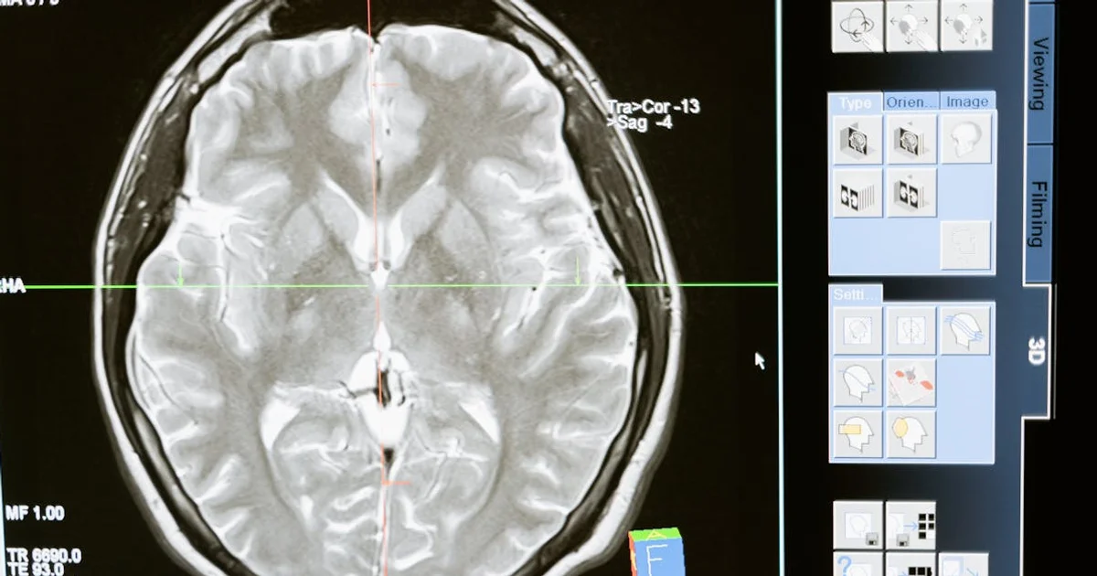
The pineal gland is a small endocrine structure in the center of the brain. Its most recognized output is melatonin — the hormone that regulates circadian rhythm and sleep-wake cycles — but the gland is also implicated in neuroendocrine aging, antioxidant signaling, and the coordination of seasonal and reproductive cycles. As people age, melatonin output from the pineal gland declines measurably, and researchers in the bioregulator field have proposed that short signaling peptides derived from pineal tissue could partly compensate for that functional loss.
In practice, Pinealon is studied as a neuroprotective agent — one that may reduce oxidative stress in neuronal tissue, support cognition, and interact with the same neuroendocrine pathways the pineal gland governs. The research is primarily preclinical (cell culture and rodent models), with a smaller number of Russian-language clinical publications. It is not approved by the FDA or EMA for any therapeutic indication, and human trial data in peer-reviewed English-language journals remains limited. Anyone curious about Pinealon should approach the literature with that context clearly in mind.
The Biology: What Pinealon Is Thought to Do
The proposed mechanism centers on neuroprotection. In cell and animal models, Pinealon has been studied for its ability to reduce neuronal apoptosis (programmed cell death) under oxidative or hypoxic stress — conditions that accelerate in aging brains. A 2016 study published in Rejuvenation Research by Khavinson and colleagues examined short peptides including Pinealon in retinal cell models and found reduction in markers of oxidative damage. The authors proposed that the peptide interacts with DNA-binding domains in a sequence-specific way, potentially influencing gene expression in neural and retinal tissue.
A key feature of all peptide bioregulators in this family is that they are short enough to cross biological barriers relatively easily and are thought to act as epigenetic modulators — influencing which genes get expressed in target tissue rather than acting like a receptor agonist or enzyme inhibitor the way most pharmaceutical drugs do. That mechanism is interesting scientifically, but it also means the downstream effects are diffuse and hard to measure with a single biomarker. In plain terms: there is no single blood test that directly tracks what Pinealon is doing.
Research from the same St. Petersburg group in animal models has linked Pinealon to improvements in learning and memory under stress conditions, reduced free-radical accumulation in brain tissue, and partial stabilization of circadian hormonal rhythms in aged animals. A 2012 paper in the Bulletin of Experimental Biology and Medicine reported cognitive benefits in aged rats following administration of pineal-derived peptide preparations. These findings are directionally interesting, but replication by independent research groups in controlled human trials has not yet been published in major English-language peer-reviewed literature.
The Melatonin Connection: Why Circadian Biology Matters for Labs
Melatonin is the pineal gland's most measurable output, and its relationship to Pinealon is worth understanding before you consider which labs to look at. Serum or salivary melatonin can be tested, though it is not on most standard panels. More practically, the downstream effects of circadian disruption do show up in routine bloodwork.
Chronic circadian disruption — which correlates with impaired pineal function — is associated with elevated fasting glucose, higher cortisol patterns, worsened lipid profiles, and elevated inflammatory markers like high-sensitivity C-reactive protein (hsCRP). A large review in Endocrine Reviews documented these metabolic consequences of disrupted circadian timing. This is relevant context: if someone is exploring Pinealon because of interest in sleep quality or cognitive aging, the metabolic markers that reflect circadian health are also the ones worth tracking over time.
So on your lab sheet, the signal of interest is not a single Pinealon-specific number — it is a cluster of markers that reflect neuroendocrine and metabolic health across repeated draws.
Which Biomarkers Are Most Relevant to Monitor?
Given the neuroprotective and neuroendocrine framing of Pinealon research, the following markers are the most logical to track for someone interested in monitoring their brain-aging or circadian biology profile. None of these directly measure Pinealon's activity — they reflect the biological territory Pinealon research operates in.
Cortisol and the HPA Axis
The hypothalamic-pituitary-adrenal (HPA) axis — the hormonal stress-response system — interacts closely with circadian timing and melatonin. Cortisol, the primary output of the adrenal glands, should peak in the morning and trough at night in a healthy circadian pattern. Serum cortisol is typically measured in the morning (roughly 8 a.m.) when levels are highest. A morning cortisol test gives a snapshot; patterns across multiple draws reveal whether the axis is functioning normally. Reference ranges vary by lab and timing, typically 6–23 mcg/dL in the morning. Disruption in the cortisol curve is one of the clearest measurable signs that circadian biology is under stress — which is directly relevant if pineal function is of interest.
IGF-1 (Insulin-Like Growth Factor 1)
IGF-1 is the primary mediator of growth hormone (GH) activity in the body — when GH is released from the pituitary gland, it travels to the liver and triggers IGF-1 production. Because GH release is pulsatile and hard to measure directly, IGF-1 serves as the stable, day-to-day surrogate. Pinealon is not a GH secretagogue like GHRP-2 or Ipamorelin, but the GH-IGF-1 axis declines with aging in parallel with pineal function, and the bioregulator research tradition that includes Pinealon frequently cites IGF-1 as part of the neuroendocrine aging picture. Tracking IGF-1 alongside cortisol gives a broader view of where someone's endocrine biology sits relative to age-matched norms. Ranges are strongly age-dependent — a level normal for a 30-year-old is low for a 20-year-old — so always compare against age-specific reference ranges, which your lab report should include.
For more on how the GH-IGF-1 axis works in the context of peptide use, see the Tesamorelin, IGF-1, and the Labs Worth Tracking explainer.
Fasting Glucose and HbA1c
Fasting glucose (the blood sugar level after an overnight fast) and HbA1c (glycated hemoglobin — a 2–3 month average of blood sugar control) are sensitive to circadian disruption and sleep quality. The pineal gland's melatonin output directly modulates insulin secretion; melatonin receptor variants have been linked to type 2 diabetes risk in genome-wide association studies, as documented in a landmark Nature Genetics paper. People exploring Pinealon for sleep or cognitive aging reasons should have a baseline fasting glucose and HbA1c on record. Normal fasting glucose is typically 70–99 mg/dL; HbA1c below 5.7% is considered non-diabetic range by most major labs.
Lipid Panel (Total Cholesterol, LDL, HDL, Triglycerides)
Lipids are worth including in any baseline panel because the neuroendocrine aging picture connects to cardiovascular risk, and cholesterol metabolism is substrate for steroid hormone synthesis — including the sex hormones and cortisol. Triglycerides in particular are sensitive to sleep disruption and circadian misalignment. Tracking total cholesterol, LDL, HDL, and triglycerides across visits gives you a metabolic fingerprint that sits alongside any neuroprotection-oriented protocol.
hsCRP (High-Sensitivity C-Reactive Protein)
High-sensitivity C-reactive protein is a sensitive marker of low-grade systemic inflammation — the kind that does not cause symptoms but is associated with accelerated aging and neurodegeneration risk over long periods. It is measured in mg/L; values below 1.0 mg/L are associated with lower cardiovascular risk, 1.0–3.0 mg/L is intermediate, and above 3.0 mg/L signals higher inflammatory burden. Because Pinealon's proposed benefits include antioxidant and anti-inflammatory effects in neural tissue, hsCRP is a reasonable proxy marker for the inflammatory context in which such effects would theoretically operate. It is also inexpensive to add to a standard panel.
TSH and Free Thyroid Hormones
Thyroid-stimulating hormone (TSH) and the free thyroid hormones (free T3 and free T4) are part of the neuroendocrine axis that regulates energy, cognition, and metabolic rate. Thyroid function interacts with sleep quality and pineal melatonin signaling. TSH is typically the first marker ordered, with the standard reference range of 0.45–4.5 mIU/L on most US labs — though the "optimal" range is debated, with some functional medicine perspectives favoring 1.0–2.5 mIU/L. For someone tracking cognitive aging or neuroendocrine biology, a thyroid panel alongside cortisol and IGF-1 provides a more complete picture than any single hormone alone.
What the Research Does and Does Not Show
Honesty about the evidence base matters here. The bulk of Pinealon research originates from one research group in Russia, published in Russian-language journals and a subset of English translations. The St. Petersburg Institute work is systematic and spans decades, but it has not been widely replicated by independent groups in double-blind randomized controlled trials with large human cohorts. That is a meaningful limitation.
What the preclinical literature does reasonably consistently show is: reduction in oxidative stress markers in neuronal cell lines, neuroprotective effects in hypoxia models, and improvements in cognition-related behavioral endpoints in aged rodents. A review of peptide bioregulators in aging by Khavinson et al. in Current Aging Science situates Pinealon within the broader bioregulator hypothesis: that tissue-specific short peptides can partially restore gene expression patterns characteristic of younger tissue. The hypothesis is coherent and biologically plausible, but the quality of evidence available in English-language peer review is not yet at the standard required for clinical recommendation.
Compared to better-characterized neuropeptides like Semax — which has a larger body of Russian clinical trial publications and some independent pharmacological characterization — Pinealon's human data is thin. Anyone evaluating it should hold that distinction clearly.
Pinealon in the Context of Broader Longevity and Cognitive Aging Panels
Interest in Pinealon typically does not arrive in isolation. People exploring it are usually also thinking about circadian health, cognitive reserve, and the pace of biological aging — a cluster of concerns that the longevity-focused lab testing world addresses through several overlapping panels. The biomarkers most consistently flagged in that literature include:
Biomarkers relevant to neuroendocrine aging and cognitive health monitoring
| Biomarker | What It Reflects | Why It Matters Here |
|---|---|---|
| Cortisol (morning serum) | HPA axis output and circadian stress response | Pineal-circadian biology; stress-aging intersection |
| IGF-1 | GH axis activity and tissue anabolism | Neuroendocrine aging; age-matched decline parallel to pineal function |
| Fasting glucose / HbA1c | Blood sugar regulation and insulin sensitivity | Melatonin-insulin interaction; circadian disruption signature |
| hsCRP | Low-grade systemic inflammation | Proxy for inflammatory aging context; inexpensive add-on |
| TSH / Free T3 / Free T4 | Thyroid axis and metabolic rate regulation | Interacts with sleep and neuroendocrine signaling |
| Lipid panel (LDL, HDL, TG) | Cardiovascular and metabolic health | Steroid hormone substrate; sensitive to sleep disruption |
| Homocysteine | Methylation capacity and cardiovascular risk | Elevated levels linked to cognitive decline in aging populations |
| Serum melatonin (specialty test) | Pineal gland output directly | Not standard; useful reference point if available |
Homocysteine is worth a brief note: it is an amino acid that accumulates when methylation pathways are insufficient, and elevated levels (above 15 micromol/L is the conventional concern threshold, though some researchers flag above 10) are associated with cognitive decline and cardiovascular risk in older adults. A 2002 landmark study in the New England Journal of Medicine linked elevated homocysteine to substantially increased dementia risk. For someone tracking cognitive aging, adding homocysteine to a standard panel costs little and provides meaningful longitudinal signal.
The picture these markers form together is more useful than any one of them in isolation. A single fasting glucose reading on one morning does not tell you much. A chart of fasting glucose, HbA1c, hsCRP, and cortisol across six visits over two years — especially when overlaid with significant life events or protocol changes — tells a story worth having a conversation about with your physician.
For more on the longevity biomarker landscape, the LabHealthCharts biomarkers library covers IGF-1 and related markers, and the Pinealon peptide page outlines the on-site framing for this substance.
Tracking Pinealon-Related Labs Over Time with LabHealthCharts
The core challenge with a research-stage peptide like Pinealon is that there is no single direct biomarker to track — so context over time becomes the only real signal you have. A cortisol result, an HbA1c, or an hsCRP value on its own tells you your number at a moment in time. Four draws across twelve months, visualized on the same timeline, start to show whether the inflammatory or metabolic environment you are managing is moving in a favorable direction.
That is where LabHealthCharts fits in. The app accepts PDFs from Quest Diagnostics, LabCorp, and most other US lab formats, uses AI-assisted extraction to pull biomarker values from those documents, and builds longitudinal charts so you can see trends across 100+ markers in one place. Instead of a folder of disconnected PDFs from different visits, you get a unified history that puts cortisol from a draw six months ago next to your most recent result, on the same axis, with the same units. For someone tracking a cluster of neuroendocrine and metabolic markers over a protocol period, that kind of visual history is not just convenient — it is the only way to see what is actually changing.
If you have results from recent labs and want to see your neuroendocrine markers visualized over time, you can upload your lab PDFs and chart them at app.labhealthcharts.com. Membership is $79/year. LabHealthCharts organizes and displays your data — it does not interpret results or provide medical guidance. That part stays with your physician.
Key Takeaways
Pinealon is a short tripeptide bioregulator studied for neuroprotection and circadian neuroendocrine support, primarily in preclinical and Russian-language clinical research. Here is what matters for anyone tracking their health in this area:
1. The evidence base is preliminary. Most data comes from one research group, primarily in animal models and cell culture. There are no large, independent, English-language randomized controlled trials. Treat Pinealon as a research-stage substance, not a clinically validated therapy.
2. No single biomarker tracks Pinealon directly. The relevant markers to monitor are a cluster that reflects the neuroendocrine and metabolic territory the research addresses: cortisol, IGF-1, fasting glucose, HbA1c, hsCRP, thyroid panel (TSH, free T3, free T4), lipid panel, and homocysteine.
3. The melatonin-insulin connection is real and measurable. If circadian biology and sleep quality are the underlying concerns, fasting glucose and HbA1c are sensitive, accessible, and well-validated markers to watch repeatedly over time.
4. Trends outperform snapshots. A single cortisol or hsCRP reading is context-poor. The same markers charted across multiple draws, aligned with changes in sleep, stress, or a protocol, give your clinician something real to work with.
5. Talk to your physician before adding any peptide or adjusting your monitoring panel. The biomarker list here is educational; individual decisions about what to test and how to interpret results belong with a qualified provider who knows your full history.